Figure 1 from Pathologic and physiologic phimosis: approach to the
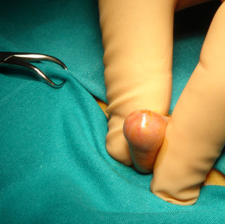
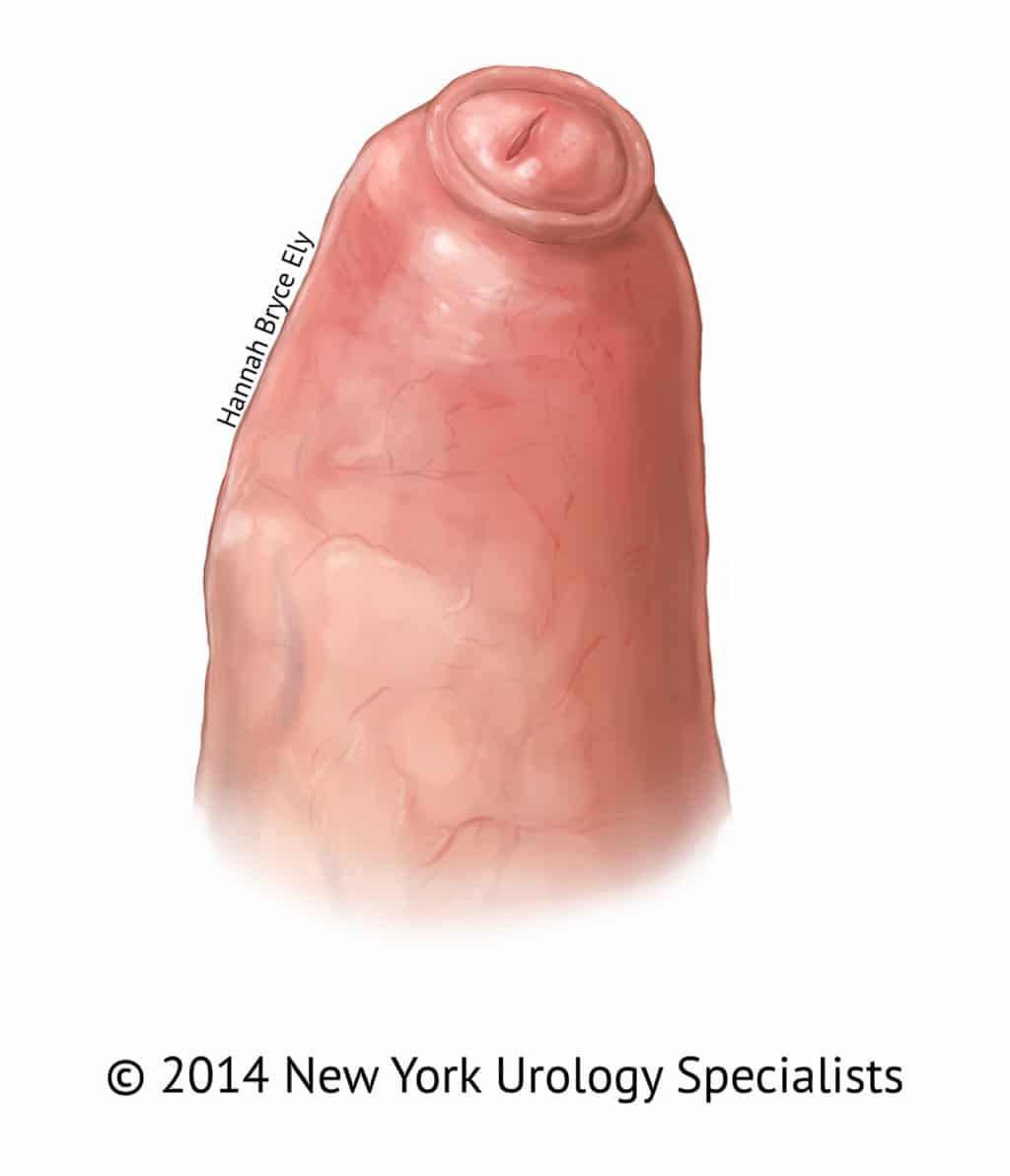
Figure 1. Tight preputial orifice on retraction of foreskin: A) Skin at preputial outlet is healthy with no scarring, and the inner preputial mucosa is starting to evert through the outlet. With physiologic phimosis, the preputial outlet is always closed and one cannot see the glans unless the foreskin is retracted, as the examiner has done in the photograph. B) In many cases of pathologic phimosis, the glans and meatus are visible without any attempt at retraction, as the scarred ring holds the preputial outlet open. There is no inner mucosal eversion through the outlet. - "Pathologic and physiologic phimosis: approach to the phimotic foreskin."
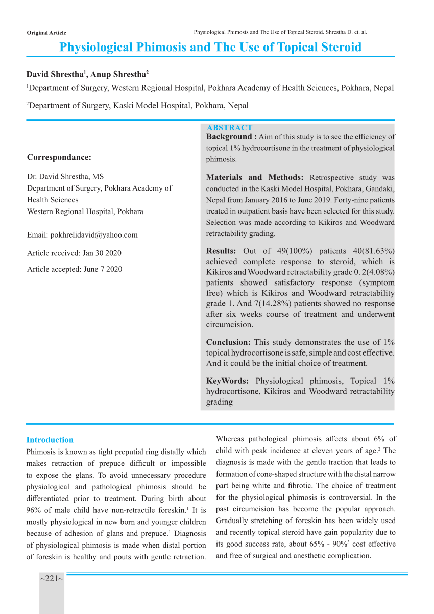
PDF) Physiological Phimosis and The Use of Topical Steroid

Figure 1, Pathologic and physiologic phimosis

CCM3 Loss-Induced Lymphatic Defect Is Mediated by the Augmented

A primary care update to circumcision
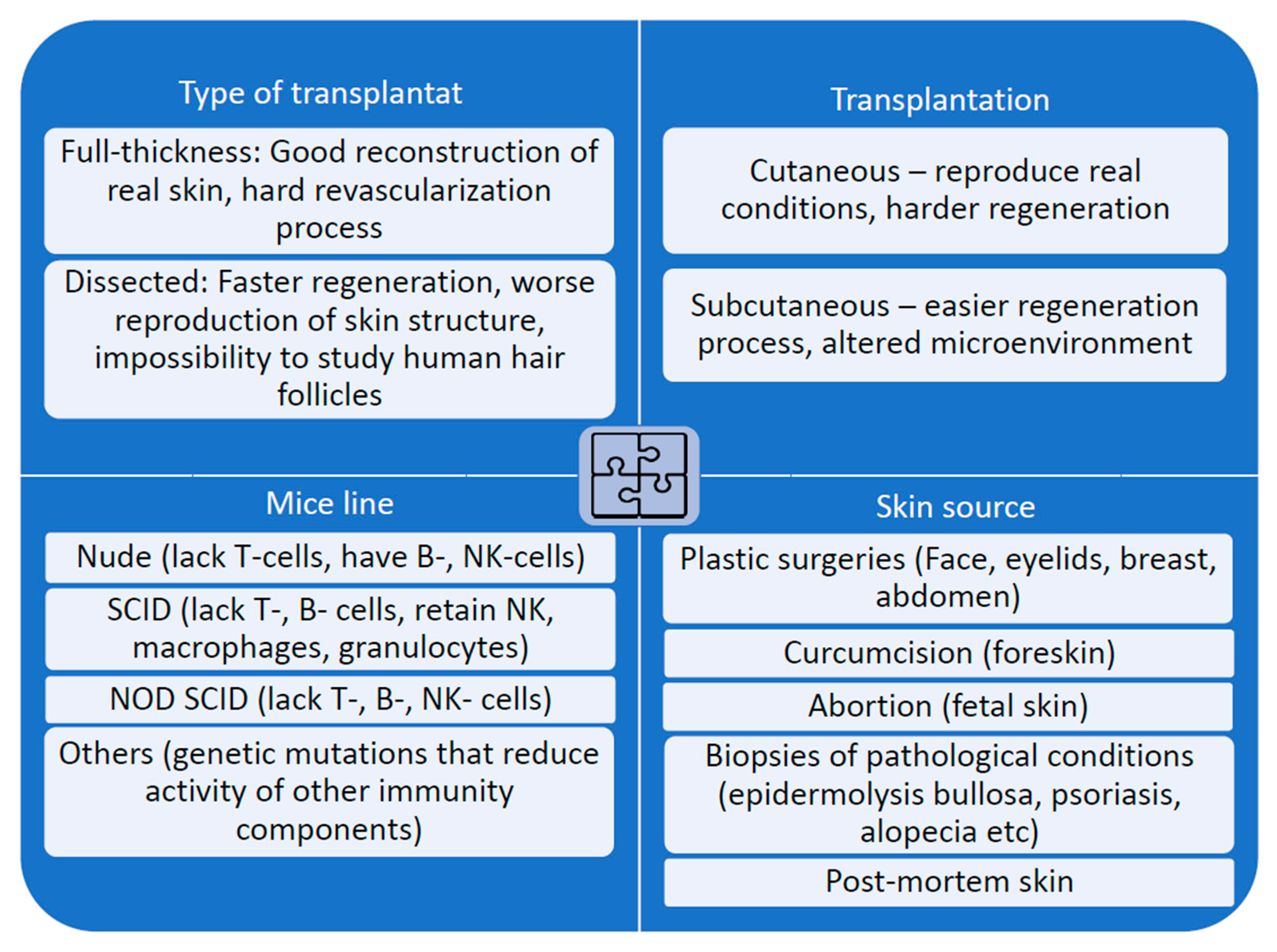
IJMS, Free Full-Text

Figure 1 from PENILE TUBERCULOSIS PRESENTING AS PHIMOSIS

Full article: Foreskin Analysis of Circumcised Boys With and

Current Approaches of Preservation of Cells During (freeze

Strategies for targeting senescent cells in human disease
Biomedicines, Free Full-Text

Projects

A primary care update to circumcision

A primary care update to circumcision
Importance of Identifying Physiological Preputial Adhesion and

Gene therapy to enhance angiogenesis in chronic wounds: Molecular